EU Funded Careioca Projet Lead to New High-Speed Ex-Vivo and Endoscopic Optical Imaging Systems for Real-Time Cancer Diagnosis Showing High Potential
PAris, France, May 20, 2016 (Newswire.com) - Cancer diagnosis relies on the long and complex histology process. During surgery, there is currently no real- time guidance at the histology level, leading to up to 40% of reoperations. Along the cancer care workflow, in particular for tumor biopsy and excision, current existing preoperative and intraoperative imaging techniques fail to perform a reliable in situ diagnosis. Moreover, the complete pathological diagnosis, often based on histology slides preparation, is usually only available after a few days. For these reasons a significant number of patients need to undergo secondary biopsy or surgery.
The CAReIOCA project combined the latest advances in CMOS camera technology and optical biopsy imaging: Full-Field Optical Coherence Tomography (FFOCT). This association resulted in the development of two innovative optical imaging tools:
- a high-speed FFOCT microscope for the non-destructive quality control of biopsies ex vivo within minutes,
- an FFOCT endoscope for in vivo surgery and biopsy guidance at the cellular level.
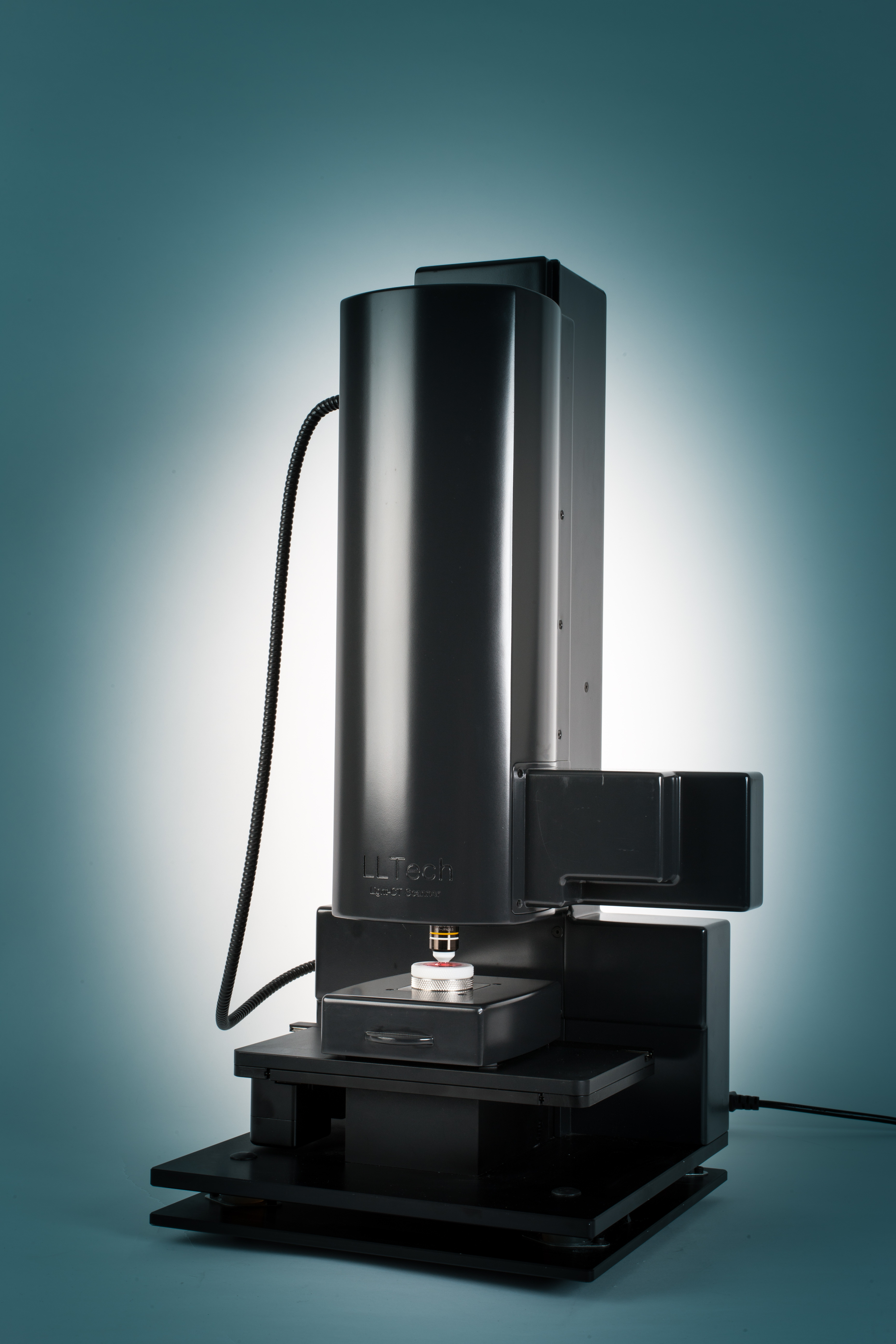
The collaboration of LLTech, specialized in the design and fabrication of FFOCT imaging devices, with CMOSIS and Adimec, who are experts in sensor and camera design, allowed breaking a major technological issue in FFOCT imaging through the development of a custom-made camera that offers a tenfold higher sensitivity than conventional cameras, at 1 kHz frame rate. This camera was integrated in the FFOCT devices, multiplying by 5 their acquisition speed. Besides, the design of a new FFOCT system compatible with a probe and the efforts made on miniaturization gave rise to the first FFOCT endoscope.
The two FFOCT devices were evaluated at Leiden University Medical Center (LUMC, Leiden Netherlands) and Gustave Roussy Institute (Villejuif France) on breast cancer and head and neck cancer and provided very good results compared to standard histology and frozen section techniques. LUMC also performed promising FFOCT images on ovarian tissue, showing enough readable information without any tissue damaging to envision the introduction of FFOCT imaging in the clinical workflow of ovarian tissue auto-transplantation. FFOCT endoscopy performed on head and neck biopsies at Gustave Roussy Institute revealed its potential of in situ imaging of small structural features within the tissues.
The project activities included the publication of 7 papers and the outcomes were disseminated via 3 exhibitions and 24 participations to international conferences.
CAReIOCA successfully evaluated three technological foregrounds – the high-speed camera, the CMOS sensor and the FFOCT microscope – as almost ready for market launch (planned in 2016 and 2017). Although the camera and sensors were specified under the FFOCT imaging requirements, other interferometry systems could benefit from them in the medical or industrial sectors. The clinical exploration conducted during the CAReIOCA project enhanced the optical biopsy knowledge on several applications and fed the trust in the technology of future clinical users.
About LLTech
LLTech is a privately owned company founded in 2007. LLTech vision is to become the leader in real time, safe, non-destructive optical biopsies and cellular level tissue imaging for research and clinical applications (micro biopsies assessment, surgical margins...). LLTech vision is to provide optical systems that will provide intra operative imaging systems that will allow real time assessment and diagnoses of cancer, thus replacing techniques such as frozen sections. For more information, go to www.lltechimaging.com.
About Leiden University Medical Center (LUMC)
LUMC is a university medical center for research, instruction and patient care. Its unique research practice, ranging from pure fundamental medical research to applied clinical research, places LUMC among the world top. With more than 7000 staff members, the LUMC focuses on top clinical and highly specialized care. A considerable portion of the research focuses on the translation from fundamental research to its use in patient care (from bench to bedside and vice versa). Within the LUMC, the image guided surgery group is a unique collaboration between the medical, biological, chemical and technological groups to bring new technologies to the surgical theatres. For more information, go to www.lumc.nl.
About Institut Gustave Roussy (IGR)
As a European leader in cancer, IGR treats patients with any type of cancer, at any stage and is expert in rare cancers and complex treatments. With its 3000 staff (researchers, teachers, doctors and care providers) IGR integrates three founding principles: research, care and teaching.
Expert in complex tumors, IGR has centered its specificity on therapeutic innovation (2800 students and professionals trained in 2014). IGR involves medical oncology, chemotherapy, radiotherapy, surgery, interventional radiology and reconstructive surgery. 30% of the patients are included in a biomedical research program (369 clinical trials in 2014). For more information, go to www.gustaveroussy.fr.
The imaging and cytometry platform (PFIC) is one of the 7 technical facilities of IGR, supporting basic and clinical research programs in oncology. The PFIC is a service and R&D platform at the interface of basic, translational and clinical research. PFIC is a partner in several pre-clinical programs and national and international clinical trials, aimed at the development and transfer of non-invasive photonic imaging at high resolution in small animals and patients.
About CMOSIS
CMOSIS is a pure-play supplier of standard, off-the-shelf as well as custom area and line scan CMOS image sensors. CMOSIS' imagers feature global and rolling shutter, low noise, high dynamic range and high frame rates through high-speed on-chip ADC and digital interfaces. CMOSIS' image sensors serve a broad range of applications for diverse markets including machine vision, medical, broadcast, traffic, scientific and photography imaging. The CMOSIS offer further includes miniature camera modules for endoscopy-like applications.
CMOSIS offers innovative turnkey image sensor solutions from specification and design, over prototyping and product qualification, to volume production in a one-stop-shop model.
CMOSIS operates from Belgium, Germany, Portugal and the USA and employs more than 115 people. CMOSIS is a member of the ams group. ams sensor solutions take sensing to the next level by providing a seamless interface between humans and technology and enable our customers to create highly differentiated products that are smarter, safer, easier to use and more eco-friendly. ams develops high-performance solutions for the most challenging applications in sensors, sensor interfaces, power management and wireless. For more information, go to www.cmosis.com.
About Adimec
Adimec specializes in the development and manufacturing of high-performance cameras that meet the application-specific requirements of key market segments, including machine vision, healthcare, and global security. Founded in 1992, the company partners with major OEMs around the world to facilitate the creation of industry-leading cameras. Its products meet a wide range of performance, size, cost, interface and application requirements and its Adimec True Accurate Imaging® technology provides new levels of precision and accuracy to vision systems. Adimec has offices around the world focused on creating customer value and satisfaction through local, personalized support. For more information, go to www.adimec.com.
Source: LLTECH